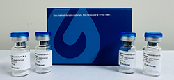
Product Description |
Murine leukemia viruses (MuLVs) are retroviruses commonly used as gene
transfer vectors in cell and gene therapy due to their capacity to integrate
into the host genome. It is important to ensure the quality and consistency of
MuLV particles during production by using accurate physical titer measurement
techniques, such as p30 antigen detection. For purification
and production of MuLVs, MuLVs titration is an essential characterization
procedure for routine quality control, lot release, and purification process
optimization. GenScript MuLV Titer p30 ELISA Kit (Cat. No. L01041) is a sandwich
ELISA kit that accurately and precisely measures the physical titer of MuLVs by
detecting the MuLV core protein p30 during viral vector production. This kit
facilitates the ease, accuracy, and reproducibility of MuLVs titration. |
LLOQ |
0.3125 ng/mL or 2.82E+06 VP/mL |
ULOQ |
5 ng/mL or 4.51E+07 VP/mL |
Assay Working Ranges |
0.3125 to 5 ng/mL (2.82E+06~4.51E+07 VP/mL) |
Kit Contents |
Component |
Quantity/Size |
Part No. |
Capture Plate |
1 plate |
X1-80 |
Standard Stock |
1 vial (50 μL) |
X1-10 |
Detection Antibody |
1 vial (12 mL) |
X1-20 |
Streptavidin-HRP |
1 bottle (12 mL) |
X1-30 |
Sample Dilution Buffer |
1 bottle (60 mL) |
X1-60 |
20× Wash Solution |
1 bottle (60 mL) |
X1-70 |
Lysis Solution |
1 bottle (10 mL) |
X1-A0 |
TMB Solution |
1 bottle (12 mL) |
A1-40 |
Stop Solution |
1 bottle (6 mL) |
A1-50 |
Plate Sealer |
2 pieces |
/ |
|
Storage |
The
unopened kit is stable for at least 12 months from the date of manufacture at
2°C to 8°C, and the opened kit is stable for up to 1 month from the date of
opening at 2°C to 8°C. |
Assay Principle |
MuLV Titer p30 ELISA Kit is
based on the sandwich ELISA method. When standards or samples are added to the
capture plate coated with anti-MuLV p30 monoclonal antibody, MuLV particles in
the samples are captured on the plate. The detection antibody, also an
anti-MuLV p30 monoclonal antibody conjugated with biotin, is then added to
interact with the MuLV particles bound to the plate. Streptavidin-HRP is added
to interact with the biotin-conjugated detection antibody. After washing steps,
TMB Solution is added
to the plate and color development is stopped by the addition of stop solution. Application of the Stop Solution
results in the color changing from blue to yellow. The color intensity can be
read at 450 nm and 650 nm by a microplate reader. The standard curve can be used to accurately
quantify the amount of MuLV in
the samples. |
Reference |
1. Renner TM, Tang VA, Burger D, Langlois MA. Intact Viral Particle Counts
by Flow Virometry Insight into Moloney Murine Leukemia Virus Infectivity and
Genome Packaging. J Virol. 2020;94(2) 2. ICH: Q2(R2) Validation of Analytical
Procedures 3. ICH: M10
Bioanalytical Method Validation and Study Sample Analysis |
Typical MuLV p30 and MuLV standard curves fitted with a four-parameter logistic model. Each standard was analyzed in duplicate, with an R-squared value of 1.
For research use only. Not intended for human and animal
therapeutic or diagnostic use.