目录产品 » 稳定细胞系 » Human Recombinant Vasoactive Intestinal Polypeptide Receptor Stable Cell Line
CHO-K1/PAC1/Gα15 Stable Cell Line

Figure 2. Dose dependent stimulation of intracellular cAMP accumulation upon treatment with PACAP in CHO-K1/PAC1/Gα15 cells. d2 acceptor fluorophore -labeled cAMP (Cat. No. 62AM4PEC; Revvity) and intracellular cAMP in CHO-K1/PAC1/Gα15 cells competitively bind with Europium Cryptate-labeled anti-cAMP monoclonal antibody. The FRET signal decreases as the intracellular cAMP concentration rises and was measured by plate reader (Pherastar, BMG). The EC50 of PACAP on CHO-K1/PAC1/Gα15 cells was 21.91 nM.

CHO-K1/PAC1/Gα15 Stable Cell Line

Figure 1. PACAP-induced concentration-dependent stimulation of intracellular calcium mobilization in CHO-K1/PAC1/Gα15 cells. The cells were loaded with Calcium-4 (Cat. No. R8142; Molecular Devices) prior to stimulation with PAC1 agonist, PACAP. The intracellular calcium change was measured by FLIPRTETRA. The relative fluorescent units (RFU) were recorded and normalized to plot against the log of the cumulative doses of PACAP (mean ± SEM, n = 3). The EC50 of PACAP on CHO-K1/PAC1/Gα15 cells was 68.90 nM.

Notes:
EC50 value is calculated with four parameter logistic equation:
Y=Bottom + (Top-Bottom) / (1+10^ ((LogEC50-X)*Hill Slope))
X is the logarithm of concentration. Y is the response.
Y is RFU and starts at Bottom and goes to Top along a sigmoid curve.

CHO-K1/PAC1/Gα15 Stable Cell Line

Recombinant CHO-K1 cells stably overexpress human pituitary adenylate cyclase-activating polypeptide type I receptor (PAC1) on the surface and contain high levels of G protein Gαq and Gαs to couple with the receptor in downstream signaling pathways.
M00203
询价

联系我们
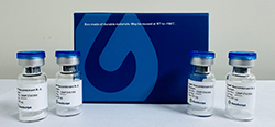
Product Description Recombinant CHO-K1 cells stably overexpress human pituitary adenylate cyclase-activating polypeptide type I receptor (PAC1) on the surface and contain high levels of G protein Gαq and Gαs to couple with the receptor in downstream signaling pathways.
Applications Calcium flux assay, cAMP accumulation assay.

Expressed Gene NCBI reference sequence NM_001118; no expressed tags
Target Protein NP_001109
Host Cell CHO-K1
Size Two vials of frozen cells (>1×10⁶ per vial in 1 mL)
Storage Store cells in liquid nitrogen immediately upon receipt. Thaw and recover cells within one year from the date received.
Mycoplasma Status Negative. The mycoplasma test was performed with MycoAlert™ PLUS Mycoplasma Detection Kit (Cat. No. LT07-318, Lonza).
Stability Stable through more than 16 passages with no significant changes in assay performance or expression profile.

Culture Properties Adherent
Freeze Medium 45% Ham’s F-12K (Kaighn’s) (Cat. No. 21127, Life Technologies), 45% FBS (Cat. No. 10099-141, Life Technologies), 10% DMSO (Cat. No. D2650, Sigma)
Complete Growth Medium Ham’s F-12K (Kaighn’s), 10% FBS
Culture Medium Ham’s F-12K (Kaighn’s), 10% FBS, 400 μg/ml Geneticin (Cat. No. 10131-035, Life Technologies), 100 μg/ml Hygromycin B (Cat. No. 10687010, Life Technologies)

Gene Synonyms ADCYAP1R1, PAC1, PAC1R, PACAPR, PACAPRI
Background The vasoactive intestinal peptide/pituitary adenylate cyclase activating polypeptide (VPAC) receptors are divided into at least three types: PAC1, VPAC1, and VPAC2. Several splice variants of PAC1 result in proteins that differ at the N-terminus and the third intracellular loop. These variants differ in their affinities for PACAP and abilities to activate Gq and Gs. High level of PAC1 expression is observed in the CNS and the adrenal medulla.

  • CHO-K1/PAC1/Gα15 Stable Cell Line
  • CHO-K1/PAC1/Gα15 Stable Cell Line

    Figure 2. Dose dependent stimulation of intracellular cAMP accumulation upon treatment with PACAP in CHO-K1/PAC1/Gα15 cells. d2 acceptor fluorophore -labeled cAMP (Cat. No. 62AM4PEC; Revvity) and intracellular cAMP in CHO-K1/PAC1/Gα15 cells competitively bind with Europium Cryptate-labeled anti-cAMP monoclonal antibody. The FRET signal decreases as the intracellular cAMP concentration rises and was measured by plate reader (Pherastar, BMG). The EC50 of PACAP on CHO-K1/PAC1/Gα15 cells was 21.91 nM.

  • CHO-K1/PAC1/Gα15 Stable Cell Line
  • CHO-K1/PAC1/Gα15 Stable Cell Line

    Figure 1. PACAP-induced concentration-dependent stimulation of intracellular calcium mobilization in CHO-K1/PAC1/Gα15 cells. The cells were loaded with Calcium-4 (Cat. No. R8142; Molecular Devices) prior to stimulation with PAC1 agonist, PACAP. The intracellular calcium change was measured by FLIPRTETRA. The relative fluorescent units (RFU) were recorded and normalized to plot against the log of the cumulative doses of PACAP (mean ± SEM, n = 3). The EC50 of PACAP on CHO-K1/PAC1/Gα15 cells was 68.90 nM.

    Notes:
    EC50 value is calculated with four parameter logistic equation:
    Y=Bottom + (Top-Bottom) / (1+10^ ((LogEC50-X)*Hill Slope))
    X is the logarithm of concentration. Y is the response.
    Y is RFU and starts at Bottom and goes to Top along a sigmoid curve.


For research use only. Not intended for human or animal clinical trials, therapeutic or diagnostic use.


喜欢新升级的网站吗?

讨厌

不喜欢

一般

喜欢

非常喜欢

*