Product Description |
Recombinant CHO-K1 cells stably overexpress human protease-activated receptor 2 (PAR2) on the surface and contain high levels of G protein Gαq to couple with the receptor in downstream signaling pathways. |
Applications |
Calcium flux assay. |
Expressed Gene |
NCBI reference sequence NM_005242; no expressed tags |
Target Protein |
NP_005233 |
Host Cell |
CHO-K1 |
Size |
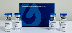
Two vials of frozen cells (>1×10⁶ per vial in 1 mL) |
Storage |
Store cells in liquid nitrogen immediately upon receipt. Thaw and recover cells within one year from the date received. |
Mycoplasma Status |
Negative. The mycoplasma test was performed with MycoAlert™ PLUS Mycoplasma Detection Kit (Cat. No. LT07-318, Lonza). |
Stability |
Stable through more than 16 passages with no significant changes in assay performance or expression profile. |
Culture Properties |
Adherent |
Freeze Medium |
45% Ham’s F-12K (Kaighn’s) (Cat. No. 21127, Life Technologies), 45% FBS (Cat. No. 10099-141, Life Technologies), 10% DMSO (Cat. No. D2650, Sigma) |
Complete Growth Medium |
Ham’s F-12K (Kaighn’s), 10% FBS |
Culture Medium |
Ham’s F-12K (Kaighn’s), 10% FBS, 200 μg/ml Zeocin (Cat. No. R250-01, Life Technologies), 100 μg/ml Hygromycin B (Cat. No. 10687010, Life Technologies) |
Gene Synonyms |
GPR11, F2RL1 |
Background |
Protease-activated receptors (PAR) are a subfamily of G-protein coupled, seven-transmembrane domain receptors, which are cleaved within the aminoterminal exodomain by certain serine proteases at a specific peptide bond. Trypsin and mast cell tryptase, and more recently, the activated coagulation factors VIIa and Xa, have been identified as serine proteases able to activate mammalian PAR-2. As already indicated, PAR-2 is believed to be involved in inflammation. This role for PAR-2 implies that elastase and cathepsin G would paradoxically display an anti-inflammatory property by disarming PAR-2. |
Figure 1. Trypsin-induced concentration-dependent stimulation of intracellular calcium mobilization in CHO-K1/PAR2/Gα15 cells. The cells were loaded with Calcium-4 prior to being stimulated with agonist glucagon. The intracellular calcium change was normalized and measured by FLIPR. The relative fluorescent units (RFU) were normalized and plotted against the log of the cumulative doses of trypsin (Mean ± SEM, n = 3). The EC50 of trypsin on this cell was 4.42 nM.
Notes:
EC50 value is calculated with four parameter logistic equation:
Y=Bottom + (Top-Bottom) / (1+10^((LogEC50-X)*Hill Slope))
X is the logarithm of concentration. Y is the response
Y is RFU and starts at Bottom and goes to Top with a sigmoid shape.
For research use only. Not intended for human or animal clinical trials, therapeutic or diagnostic use.
如您需要获取细胞系产品的说明书,请点击“Manual”,完善信息后点击“提交”。
我们会第一时间联系回复您。注意:中国地区购买的产品,说明书需要以中文站为准。
您也可拨打400-0258686转5810,或发邮件至product@genscript.com.cn联系我们。
更多产品文件类型 >>